ECG normal:

ECG normal (electrocardiograma) (onda P, segmento PR, intervalo PR,  complejo QRS, intervalo QT, segmento ST, onda T, onda U ) Vector de stock  #105838498 de ©stockdevil_666

ST elevado:

ECG de infarto de miocardio con elevación del ST (IAMCEST) y detalle del ECG  (onda P,

Presentaciones de ST elevado:

Electrocardiogram (ECG) abnormalities in the ST-segment elevation myocardial infarction (STEMI) patient. (a) Convex ST-segment elevation morphology, very common in the acute myocardial infarction (AMI) patient, (b) Obliquely straight ST-segment elevation morphology, very common in the AMI patient, (c) Concave ST-segment elevation morphology, frequently seen early in the course of the acute infarction; this ST-segment elevation morphology, however, is much less common later in course of the AMI patient, (d) The hyperacute T wave of early STEMI, usually seen in the initial 5-30 min of STEMI; with the appropriate clinical presentation, the hyperacute T wave is considered a STEMI equivalent, (e) Reciprocal ST-segment depression, more commonly known as reciprocal change, is defined as ST-segment depression on the ECG of a patient with simultaneous ST-segment elevation. Its presence, in a patient with the appropriate clinical presentation, is strongly suggestive of STEMI. Note that the concept of reciprocal change cannot be used in the setting of left bundle branch block, right ventricular paced pattern, and the left ventricular hypertrophy via voltage pattern-the ST-segment depression seen in these patterns is not considered reciprocal change. STEMI: ST-segment elevation myocardial infarction

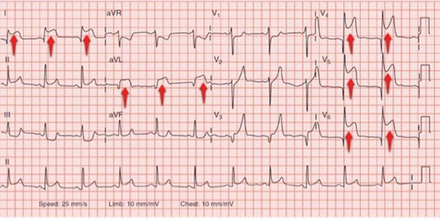
Ejemplo de ST elevado:

Segmento ST | Valoracion y Analisis del Segmento ST

Infradesnivel del ST:

ECG de infarto de miocardio sin elevación del ST (IAMSEST) y detalle del ECG (onda P, segmento PR, intervalo PR, complejo QRS, intervalo QT, depresión ST, onda T) Síndrome coronario agudo, angina de pecho

Segmento ST | Valoracion y Analisis del Segmento ST

Ejemplo infradesnivel del ST:

136 MARZO:83 abril.qxd.qxd